Optogenetics
Interested in bringing the power of Optogenetics to your patch clamping protocols?
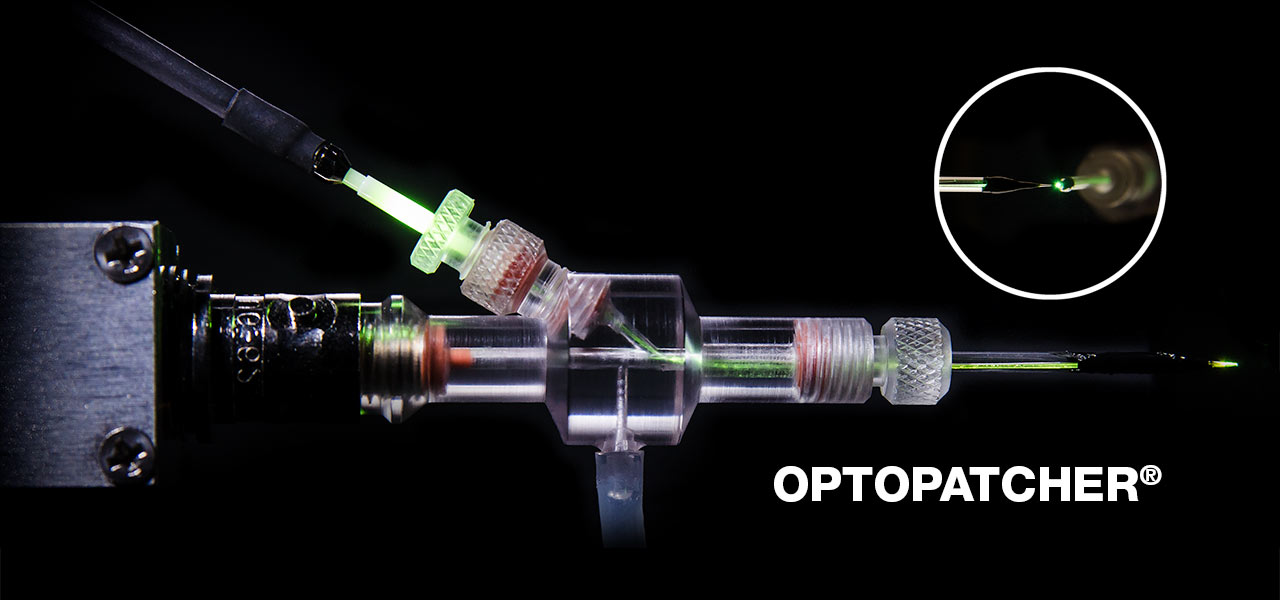
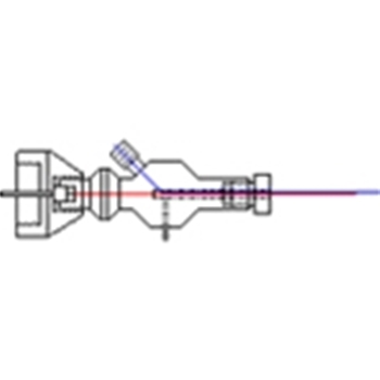
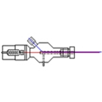
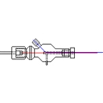
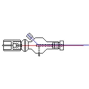
The Optopatcher® provides unmatched accuracy in applying optical stimulation to an in-vivo patch-clamp protocol. The holder houses both an optical fiber and an electrode enabling simultaneous patch-clamp recording and optogenetic activation.
This is a significant advantage in non-slice protocols, such as in-vivo recording from intact animals, whole-cell patch-clamping, LFP recording, and spike/single-unit recording.
The Optopatcher® is available with the most common connectors used on patch clamps: Axon's Threaded Collar, Axopatch and Axoclamp connectors, and standard BNC connectors used by Heka and A-M Systems. It can be ordered to accept capillary glasses with diameters between 1.2 mm and 2.0 mm. For custom diameters, please contact us.
To connect to the Optopatcher®, a compatible fiber optic cable terminating in a 1.25mm ceramic ferrule is required. We offer 1 m long cables terminating in FC/PC connectors, SMA connectors, and others.
The fiber supplied in the Optopatcher® needs to be cut to final length for proper fitting within the patch pipette. Previous work by the Optopatcher® developers determined that the fiber end did not need any special tip treatment for effective light transmission. A blunt cut non-polished fiber end could elicit biological responses provided the fiber end was positioned near the end of the pipette. We recommend that first time Optopatcher® purchasers also purchase one of our ruby blade knives (if your lab doesn't already have one).
About Optopatcher®
The Optopatcher® was developed by A-M Systems under the guidance of its inventors, Dr. Ilan Lampl and Dr. Yonatan Katz of the Weizmann Institute of Science in Israel. (J Neurosci Meth 214 (2013) 113– 117).
Each Optopatcher® Includes
- One starter fiber
- Replacement seals
- One mating sleeve for the ferrule

